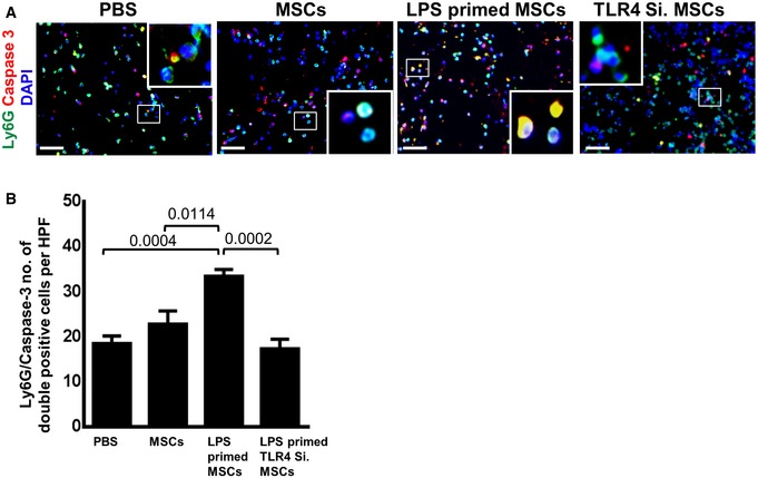
Figure EV5. A significant increase in the caspase 3 activity of neutrophils at an early stage of wound healing in wounds injected with LPS‐primed MSCs.

- Representative photomicrographs of sections from differently injected day 3 wounds stained for Ly6G+ neutrophils (green) and caspase 3 (red). Nuclei are stained with DAPI (blue). Double staining was performed for sections of day 3 wounds injected with PBS, non‐primed MSCs, LPS‐primed MSCs, and LPS‐primed TLR4‐silenced MSCs. Double‐stained cells indicate apoptotic neutrophils. To facilitate comparison, areas inside the rectangles are shown at 5× magnification in the insets. Scale bars: 50 μm.
- Quantification of Ly6G and caspase double‐positive cells on sections of differently injected day 3 wounds. A significant increase in double‐positive cells (Ly6G and caspase 3) in sections of wounds injected with LPS‐primed MSCs as opposed to wounds injected with LPS‐primed TLR4‐silenced MSCs and PBS control. Double‐positive cells were counted; statistical analysis was performed using one‐way ANOVA, and values are represented as mean ± SEM, six biological replicates.
