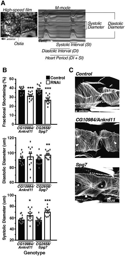
Figure 4.

Within CNV segment 12, Ankrd11 and Spg7 produce moderate alterations in heart function and structure. The Drosophila heart was used as an in vivo model system to assess the effects of candidate gene KD specifically in the heart using Hand4.2-GAL4 on function and structure (A). Hearts were filmed with a high-speed camera and analyzed using SOHA analysis, following which hearts were fixed and stained for phalloidin to demarcate filamentous actin. Within CNV segment 12, a moderate decrease in FS (top) caused largely by increased SD (bottom) was detected in Ankrd11 and Spg7 KD hearts (B). Phalloidin staining of the hearts visualized the cytoskeletal structure, whereby KD of Spg7 produced actin filament disorganization, whereas Ankrd11 KD produced minimal changes (C). Arrow heads indicate myofibrillar disorganization, whereas asterisks indicate gaps/holes in the actin filament structure. *P < 0.05, **P < 0.01 and ***P < 0.001.
