
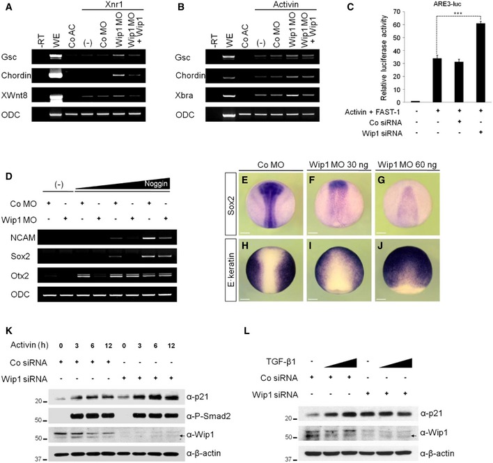
Figure 2. Depletion of Wip1 up‐regulates TGF‐β signaling.
-
A, BActivin/nodal‐induced gene responses are enhanced further in the absence of Wip1 as analyzed by RT–PCR. Animal caps in (B) were treated with activin protein (5 ng/ml). (–), no injection of Co MO, Wip1 MO, and/or Wip1 mRNA. The dose of reagent injected: Xnr1 (5 pg), Co MO (60 ng), Wip1 MO (60 ng), and Wip1 (1 ng).
-
CKnockdown of Wip1 up‐regulates the activity of activin/nodal‐responsive promoter. HEK293T cells transfected with Co siRNA (50 nM) or Wip1 siRNA (50 nM) were stimulated with activin protein (10 ng/ml) for 10 h and then subjected to luciferase assays. Data are expressed as the mean ± SEM (n = 3 biological replicates). ***P < 0.001 by unpaired Student's t‐test.
-
D–JDepletion of Wip1 promotes epidermal differentiation at the expense of neural fate. (–), no injection of Noggin mRNA. Embryos in (E–J) are shown in dorsal views with anterior to the top. The amount of MO and mRNA injected: Co MO (60 ng), Wip1 MO (60 ng for D, G, and J; 30 ng for F and I), and Noggin (5, 10, 20 pg). Scale bar, 150 μm.
-
K, LActivin or TGF‐β induction of p21 is up‐regulated upon Wip1 knockdown. HEK293T cells were transfected with Co siRNA or Wip1 siRNA and 48 h after siRNA transfection were stimulated with activin (10 ng/ml) for 3–12 h as indicated or with increasing concentration of TGF‐β1 (10, 20 ng/ml) for 3 h. t = 0 in (K) is 48 h after siRNA transfection. P‐Smad2 serves as an indicator of active activin/nodal signaling. Arrows indicate non‐specific bands.
Source data are available online for this figure.