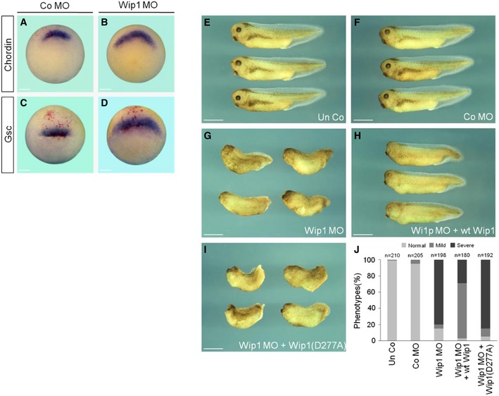
Figure 4. Mesoderm expansion in Wip1‐depleted embryos.

-
A–DKnockdown of Wip1 expands the expression of dorsal mesodermal markers. Embryos injected dorsally with Co MO (40 ng) or Wip1 MO (40 ng) were subjected to in situ hybridization at stage 10.25. Embryos are shown in dorso‐vegetal views with dorsal to the top. Scale bar, 150 μm.
-
E–IMorphological phenotypes of Wip1 morphants. Embryos were injected radially in the marginal zone with Co MO (80 ng), Wip1 MO (80 ng), wt Wip1 (1 ng), and Wip1 (D277A) mRNA as indicated and cultured to tadpole stages. Embryos are shown in lateral views with anterior to the left. Un Co, uninjected control. Scale bar, 1 mm.
-
JQuantification of the phenotypes shown in (E–I). Severe defects include microcephaly, shortened and kinked body axis, and no eye. Mild defects indicate normal body axis with malformed eyes.
