-
A
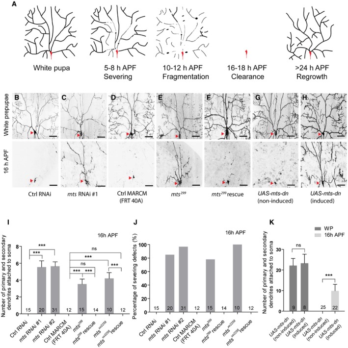
A Schematic representation of dendrite pruning in ddaC neurons.
-
B–F
Live confocal images of ddaC neurons expressing UAS‐mCD8‐GFP driven by ppk‐Gal4 at WP and 16 h APF stages. Dendrites of ctrl RNAi (B), mts RNAi #1 (C), ctrl MARCM (D), mts
299 MARCM (E), and mts
299 rescue (F) ddaC neurons at WP and 16 h APF stages. Red arrowheads point to the ddaC somas.
-
G, H
UAS‐mts‐dn ddaC neurons from animals in RU486‐induced condition (H) driven by GeneSwitch‐Gal4‐2295 exhibited normal arbors at WP stage and severe dendrite pruning defects at 16 h APF, compared to those in a non‐induced condition (G). Red arrowheads point to the ddaC somas.
-
I–K
Quantification of number of primary and secondary dendrites attached to soma and percentage of severing defects at 16 h APF.
Data information: In (I–K), data are presented as mean ± SEM from three independent experiments. One‐way ANOVA with Bonferroni test (I) and two‐tailed Student's
t‐test (K) were applied to determine statistical significance. ns, not significant; ***
P < 0.001. The number of neurons (n) examined in each group is shown on the bars. Scale bars in (B–H) represent 50 μm.
Source data are available online for this figure.

