-
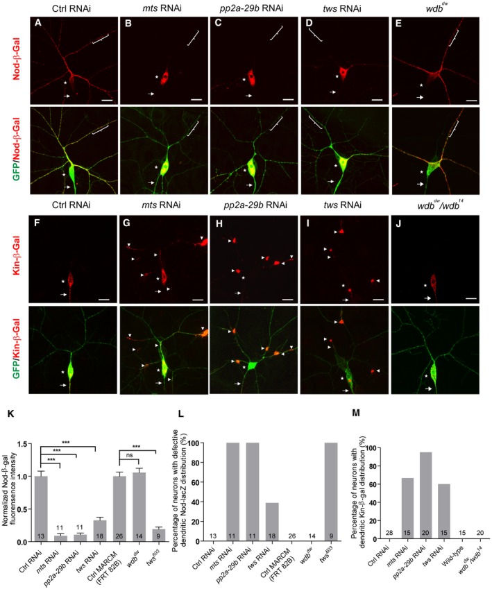
A–E
Confocal images of ddaC neurons at the wandering 3rd instar (wL3) stage immunostained for anti‐β‐galactosidase. Nod‐β‐gal signals were localized in the dendrites of the ctrl RNAi (A) and wdb
dw (E) mutant ddaC neurons; however, Nod‐β‐gal levels were strongly reduced in the dendrites and accumulated in the somas in mts RNAi (B), pp2a‐29b RNAi (C), and tws RNAi (D) ddaC neurons. ddaC somas are marked by asterisks, axons by arrows, and dendrites used for analyzing by curly brackets.
-
F–J
Kin‐β‐gal was mislocalized to the dendrites in mts RNAi (G), pp2a‐29b RNAi (H), tws RNAi (I), and wdb
dw/
wdb
14 (J) mutant ddaC neurons, compared to the ctrl RNAi neurons (F), while the localization of Kin‐β‐gal in wdb
dw/
wdb
14 mutant (J) is normal. ddaC somas are marked by asterisks, axons by arrows, and white arrowheads point to dendritic Kin‐β‐gal signals.
-
K
Quantification of normalized Nod‐β‐gal fluorescence intensity in dendrites ofddaC neurons.
-
L, M
Quantification of the percentage of neurons with defective Nod‐β‐gal and Kin‐β‐gal distribution in ddaC neurons.
Data information: In (K–M), data are presented as mean ± SEM from three independent experiments. ns, not significant; ***
P < 0.001 (one‐way ANOVA with Bonferroni test). The number of neurons (n) examined in each group is shown on the bars. Scale bars in (A–J) represent 10 μm.
Source data are available online for this figure.