FIG. 2.

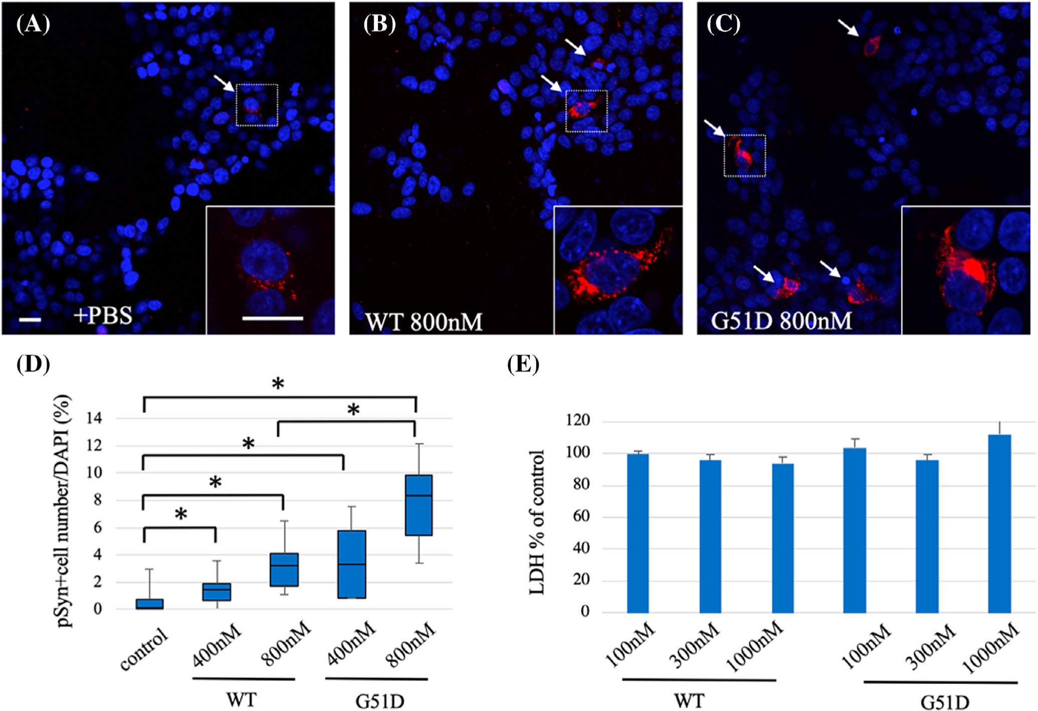
G51D α-syn fibrils promote formation of phosphorylated α-syn inclusion in mammalian cells. Representative images of dual staining of p-α-syn (red) and nuclear stain DAPI (blue) of human WT α-syn stably overexpressing SH-SY5Y cells (SH-SY5Y WT α-syn) treated with (A) PBS, (B) WT, or (C) G51D α-syn fibrils. Bottom right panels depict high-power photomicrographs of areas indicated by the squares, respectively. (D) Quantification of percentage of p-α-syn-positive cells of total cell from 6 random 20× magnification microscopic fields of view showed a higher percentage of p-α-syn-positive cells of total cell in G51D α-syn fibril-treated cells compared with WT α-syn-fibrils treated cells. (N = 4 independent experiments.) *P < 0.05, Wilcoxon signed-rank test. (E) LDH assay revealed addition of α-syn fibrils (100–1000 nM) did not induce LDH release in SH-SY5Y WT α-syn cells at 24 hours. Results are shown as mean ± SEM. Scale bar: 10 μm. α-syn, alpha-synuclein; DAPI, 4’,6-diamidino-2-phenylindole; LDH, lactate dehydrogenase; p-α-syn, phosphorylated α-syn; PBS, phosphate-buffered saline; WT, wild-type; SEM, standard error of mean.
