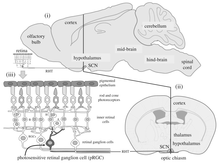
Figure 1.
The mammalian suprachiasmatic nuclei (SCN) and retina. (i) The mouse brain from the side showing the suprachiasmatic nuclei (SCN) which contains the master circadian pacemaker of mammals. The SCN receive a dedicated projection from the retina called the retino-hypothalamic tract (RHT); (ii) the frontal view of the brain shows the small-paired SCN which are located either side of the third ventricle and sit on top of the optic chiasm (where the optic nerves combine). In mice, the SCN comprises approximately 20 000 neurons, and in humans 50 000 neurons. See text for details. (iii) Retinal rods and cones convey visual information to the retinal ganglion cells (RGCs) via the second order neurons of the inner retina—the bipolar (B), horizontal (H) and amacrine (A) neurons. The optic nerve is formed from the axons of all the ganglion cells and this large nerve takes light information to the brain. A subset of photosensitive retinal ganglion cells (pRGC—shown in dark grey) can also detect light directly. The pRGCs use the blue light-sensitive photopigment, melanopsin or OPN4. Thus photodetection in the retina occurs in three types of cell: the rods, cones and pRGCs. The pRGCs also receive signals from the rods and cones, and, although not required, they can help drive light responses by the pRGCs.

