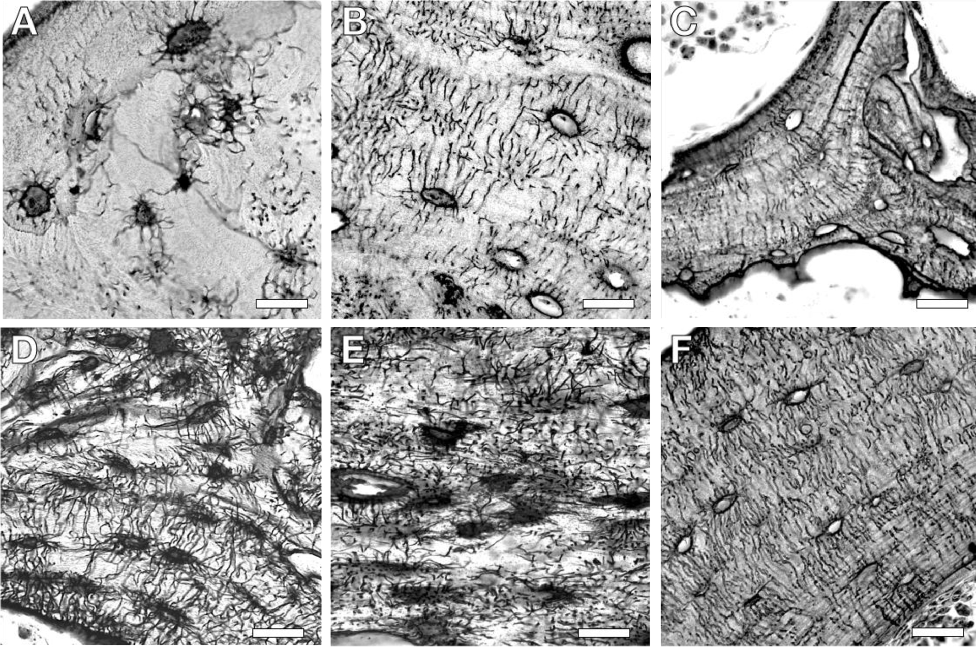
Figure 2: Diversity in the histological appearance of the lacuna-canalicular network (LCN) across species and bones.

Differences in the LCN are apparent in silver nitrate stained sections of human (A), rabbit (B), and mouse (C) trabecular bone. LCN variation also appears among bones, as shown from mouse cortical bone of the cochlea (D), mandible (E), and femur (F). Scale bar, 20 μm.
