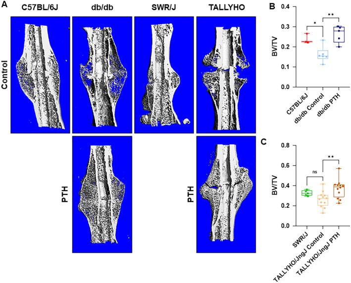
Figure 7.

μCT analysis of fracture calluses. (A) Representative images of μCT‐scanned and graphically reconstructed global images of fracture calluses at 28 days postfracture. (B,C) Assessment of the ratio of bone volume to total volume (BV/TV) of healing femora from C57BL/6J (n = 3), db/db (Control n = 6, PTH n = 5), SWR/J (n = 5), and TALLYHO/JngJ mice (Control n = 12, PTH n = 12). Db/db and TALLYHO/JngJ mice treated with hPTH (1–34) showed a significantly increased average fraction of mineralized bone (BV/TV) at 0.25 ± 0.05 (p = 0.0020) and 0.37 ± 0.09 (p = 0.0054), respectively. This is compared with untreated db/db and TALLYHO/JngJ mice groups at 0.13 ± 0.03 and 0.26 ± 0.08, respectively. C57BL/6J and SWR/J mice showed an average fraction of mineralized bone of 0.24 ± 0.02 and 0.33 ± 0.03, respectively. The difference between C57BL/6J and db/db control was significant (p = 0.0326), whereas SWR/J and TALLYHO/JngJ controls were not (p = 0.254). ns = p ≥ 0.05, *p < 0.05, **p < 0.01; one‐way ANOVA.
