
Fig. 5.
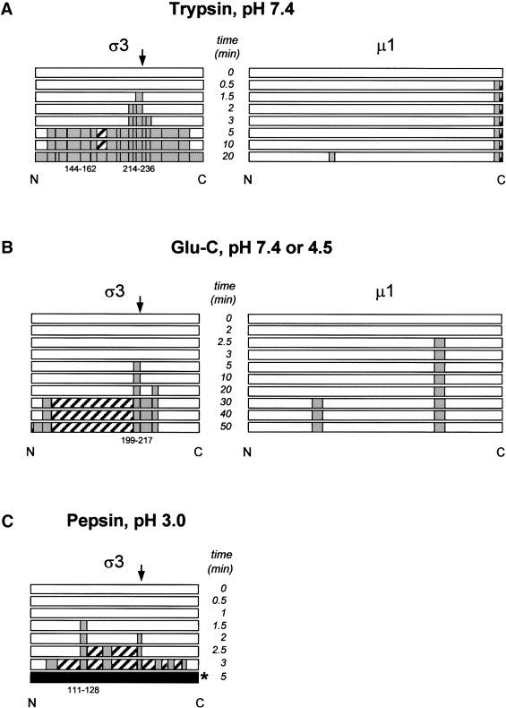
Diagrammatic representation of time-course enzymatic digests of σ3 and μ1 outer capsid proteins. (A) Time-course digestion of T1L σ3 and μ1 by trypsin at pH 7.4. (B) Time-course digestion of T1L σ3 and μ1 by Glu-C at pH 7.4 or 4.5 (results identical at both pH values). (C) Time-course digestion of T1L σ3 by pepsin at pH 3.0. White bar: undigested protein; gray bar: peptides identified by MS or MS/MS; hatched bar: nonidentified peptides; black bar: too many fragments to identify. Initial peptides cleaved are indicated with numbers below bars, with the exception of 144–162 in A (see Discussion).