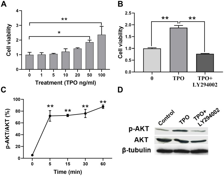
Figure 6.
TPO promoted cell proliferation and activated the PI3K/AKT signal in C17.2 cells. C17.2 cells were treated with TPO for 72 h. These data were expressed as means ± SEM. Cell viability was detected by MTT. (A) Cell viability of C17.2 cells with different TPO concentrations, n = 3. (B) Cell viability of C17.2 cells that were treated with the PI3K inhibitor (LY294002, 50 μM) prior to TPO treatment, n = 8. (C) Levels of phosphorylated AKT (p-AKT) at different time intervals. (D) Levels of AKT and p-AKT in the groups treated with TPO and TPO+LY294002. * P < 0.05, ** P < 0.01.

