Figure 6.

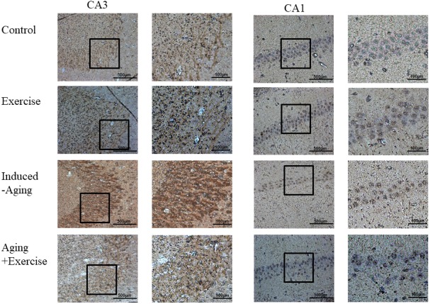
Photomicrographs of caspase 3-positive cells in the CA1 and CA3 of hippocampus. The sections were stained for caspase-3 immunoreactivity (brown).

Photomicrographs of caspase 3-positive cells in the CA1 and CA3 of hippocampus. The sections were stained for caspase-3 immunoreactivity (brown).