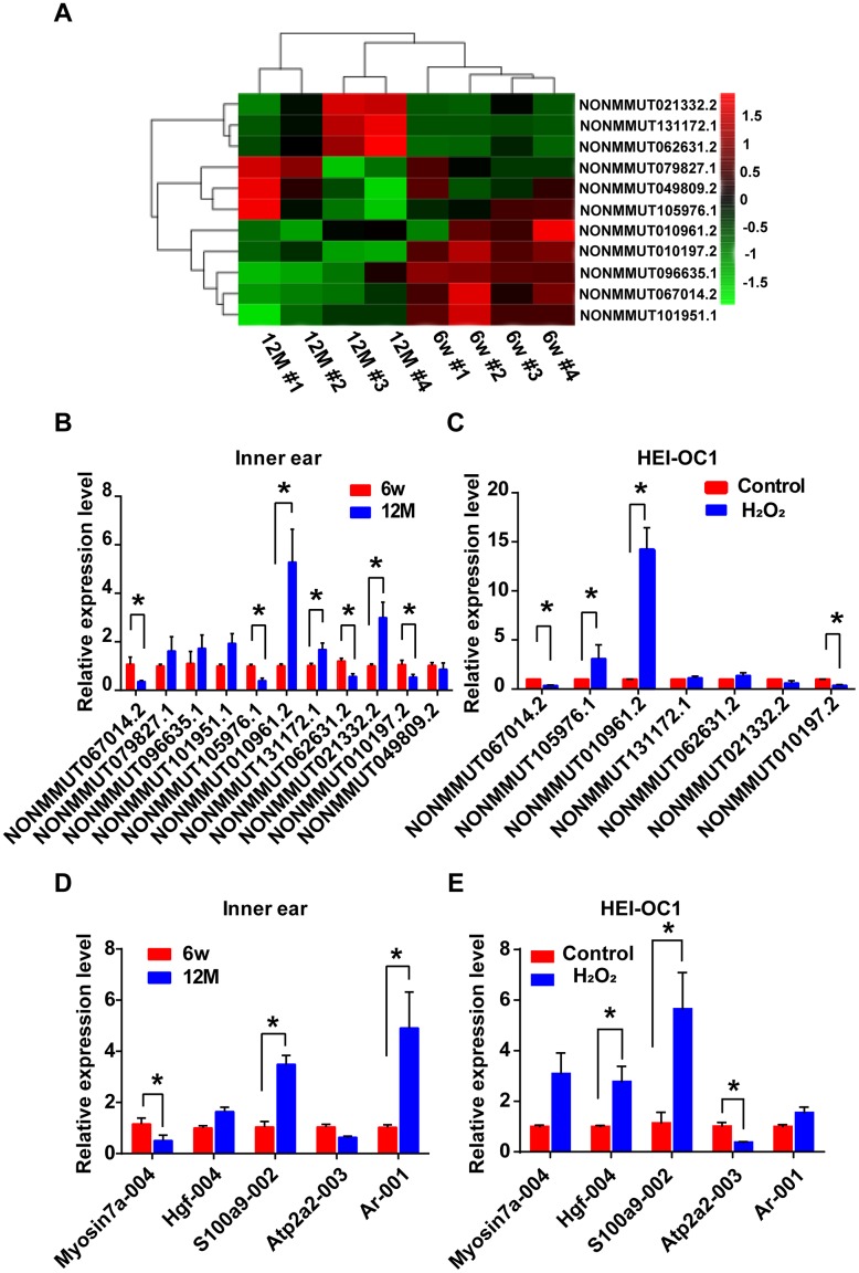
Figure 6.
Identification of differentially expressed lncRNAs and their potential target mRNAs in animal and cellular models of AHL. (A) Heat map of candidate lncRNA expression levels in six-week-old and one-year-old mice. The gradient color barcode at the top right indicates the log2 FPKM. Relative expression of the selected lncRNAs (B) and mRNAs (D) in the cochlear tissues, as detected by qRT-PCR. Tissues from the 6w mouse group were used as a control. Relative expression of the selected lncRNAs (C) and mRNAs (E) in a cellular model of AHL. Cells not treated with H2O2 were used as a control. *P < 0.05 compared to control.

