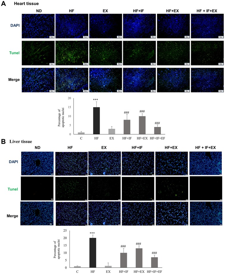
Figure 5.
IF administration during exercise show better protection from HFD induced hepatic and cardiac apoptosis in aging accelerated SAMP8 mice. TUNEL staining show the difference in TUNEL positive cells induced by HFD feeding in heart (A) and liver (B) tissues in different groups (n=6) of SAMP8 mice. The nucleus are stained in blue and the TUNEL positive apoptotic nuclei are stained in green. C: Control; HF: High-fat diet; EX: Exercise; HF+IF: High-fat diet+IF; HF+EX: High-fat diet+ Exercise; HF+EX+IF: High-fat diet+ Exercise+ IF. Scale bars, 200 μm. (Magnification, 200x). Bars indicate the mean ± SEM obtained from experiments performed in triplicate. ***P<0.001 compared with the control group, ###P<0.001 compared with the HF group.

