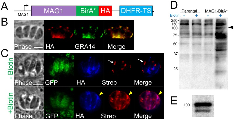
Fig 1. MAG1-BirA* localizes to the PV and biotinylates proteins in the cyst vacuole.
(A) Diagram of the construct encoding the promoter and full genomic sequence of MAG1 fused to BirA* along with a 3xHA C-terminal epitope tag. (B) IFA of MAG1-BirA* showing that the fusion protein appropriately traffics to the tachyzoite PV and colocalizes with the known protein GRA14. Scale bar: 5 μm (applicable to all panels). (C) IFA of MAG1-BirA*-expressing parasites, showing the bradyzoite PV is labeled in a biotin-dependent manner (yellow arrowheads, +Biotin row). Endogenously biotinylated apicoplasts are observed with and without biotin (white arrows, -Biotin row). Scale bar: 5 μm (applicable to all panels in C). (D) Western blot of whole-cell lysates of parental (PrugniaudΔku80Δhxgprt) and MAG1-BirA*-expressing parasites -/+ biotin. Lysates were probed with streptavidin-HRP, revealing an increase in biotinylated proteins in MAG1-BirA*-expressing parasites upon addition of biotin. The MAG1-BirA* fusion protein is predicted to be ~105 kDa (arrowhead). (E) Western blot showing that MAG1-BirA* fusion migrates to ~105 kDa as detected by antibody against HA epitope tag.

