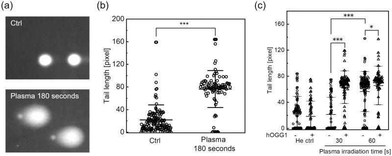
Fig 4. Strand breaks and chemical modification in A549 cells induced by He plasma jet treatment.
Cells were treated with the plasma jet, and DNA damage levels were measured using the alkaline comet assay. (a) Typical fluorescence images acquired in the alkaline comet assay without hOGG1 treatment. “Plasma 180 seconds” indicates He plasma jet irradiation for 180 seconds. “Ctrl” indicates the control experiment in which the cell suspension was exposed to He gas flow for 180 seconds at the same flow rate. (b) The tail length was measured as the DNA damage level. Each dot represents a single cell, with a minimum of 90 cells counted for each experimental condition. Data are expressed as mean±SD. Statistical significance was determined with the Student’s t-test, ***p < 0.0001. (c) The alkaline comet assay combined with human 8-oxoguanine glycosylase (hOGG1) treatment. The tail length with different plasma irradiation times and in the presence or absence of hOGG1 treatment is shown. Each dot represents a single cell, with a minimum of 80 cells counted for each experimental condition. Data are expressed as the mean±SD. Statistical significance was recognized at *p = 0.02238 and ***p < 0.0001 as determined with the Student’s t-test.

