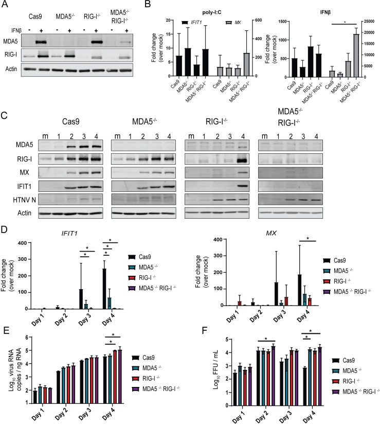
Fig 3. RLRs drive innate immune signaling and control HTNV replication.
(A) Western blotting demonstrates specific knock-outs for CRISPR-Cas9 transgenic HUV-EC-C lacking MDA5 and RIG-I. Cells were either mock-treated or treated with 10U/mL IFNβ for 24hr to induce ISG expression. (B) RT-PCR analysis of ISG transcription in HUV-EC-C KO following treatment with exogenous pI:C (1pmol) or recombinant IFNβ (10U/mL) for 18 hours (±SD). (C) Western blotting analysis for HUV-EC-C KO cells following HTNV infection (MOI 0.1) harvested for four days p.i. (D) RT-PCR of ISG expression and (E) qRT-PCR pf HTNV nucleocapsid expression in HUV-EC-C CRISPR KO cells following HTNV infection (MOI 0.1) harvested for four days p.i. (±SD) (F) Viral titer quantification from supernatants of HTNV-infected HUV-EC-C KO cells (±SD). Data shown represent three independent experiments. Statistical analysis performed with two-way ANOVA analysis, * denotes p adjusted < 0.05.

