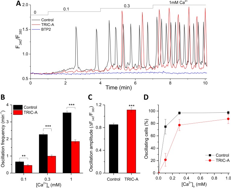
Fig 1. TRIC-A modifies the frequency and amplitude of RyR2-mediated cytosolic Ca2+ oscillations.
(A) Traces of cytosolic Ca2+-sensitive Fura-2 ratio, representing SOICR-associated oscillations in mCherry-ER-3– (control, black) or TRIC-A-mCherry–transfected (TRIC-A, red) HEK293_RyR2 cell and lack of oscillations in 3 μM BTP2-incubated (BTP2, blue) control cell. (B) Ca2+ oscillation frequency at 0.1, 0.3, and 1 mM [Ca2+]o, (C) amplitude at 1 mM [Ca2+]o, and (D) proportion of oscillating cells in TRIC-A cells (n = 88) versus controls (n = 81); **p < 0.01, ***p < 0.001; mean ± SEM values are shown. Underlying data in panels (A–D) are included in S1 Data. BTP2, N-[4-[3,5-Bis(trifluoromethyl)pyrazol-1-yl]phenyl]-4-methylthiadizole-5-carboxamide; ER, endoplasmic reticulum; Fura-2, cytosolic Ca2+-sensitive fluorescent indicator; HEK293, human embryonic kidney 293; RyR, ryanodine receptor; SOICR, store-overload–induced Ca2+ release; TRIC, trimeric intracellular cation.

