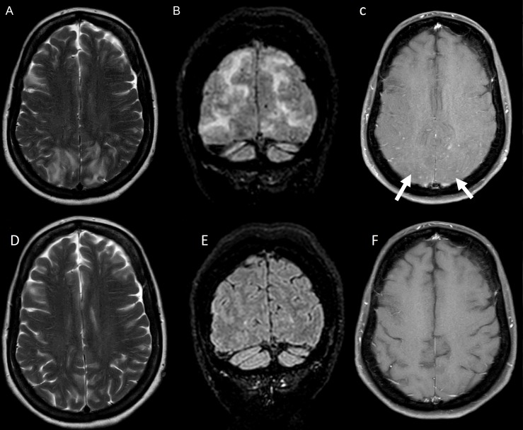
Figure 1.
MRI brain imaging performed when symptoms initially started (A–C) and at 2-week follow-up assessment (D–F). (A) and (B) Patchy bilateral symmetrical parieto-occipital subcortical T2 and fluid-attenuated inversion recovery (FLAIR) hyperintensities. (C) Subtle foci of nodular postcontrast enhancement on T1-weighted imaging. (D–F) Follow-up brain and spine imaging show resolution of initial change.

