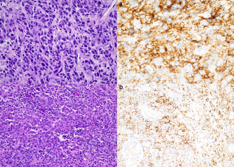
Figure 2.
Pathology and programmed cell death 1 ligand (PD-L1) analysis. (A) A liver core needle biopsy from patient 1 demonstrates a pleomorphic spindled to histiocytoid neoplasm with prominent lymphocytic infiltrate, single-cell apoptotic bodies and mitotic figures (40×, H&E). (B) Patient 1 PD-L1 263 immunohistochemistry demonstrates membranous and cytoplasmic staining of neoplastic cells (40×). (C) Patient 2 superficial soft-tissue resection shows a histiocytoid proliferation with mild-to-moderate cytologic atypia and a prominent neutrophilic and lymphocytic infiltrate (20×, H&E). (D) Patient 2 PD-L1 263 immunohistochemistry showing cytoplasmic and membranous staining of neoplastic cells (20×).

