
Fig. 2. TMEM16F CaPLSase is highly expressed in human placental trophoblasts.
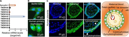
(A) qRT-PCR of TMEM16 family members in BeWo cells. All genes are normalized to glyceraldehyde-3-phosphate dehydrogenase (GAPDH) and then normalized to Syncytin-1. (B) Immunofluorescence of TMEM16F (green) in BeWo cells (upper) and the primary human placental trophoblasts from a term placenta (lower). The nuclei are stained with Hoechst (blue). (C) Immunofluorescence of TMEM16F (anti-TMEM16F, green) and nuclei (Hoechst, blue) in a human first trimester placenta villi (upper). Higher magnifications are shown in the lower panels. TMEM16F is highly expressed in both cytotrophoblasts and syncytiotrophoblasts (arrowheads). The white dotted line demarcates the basal membrane of the syncytiotrophoblast. Schematic of the maternal-fetal interface in the first trimester placental villi is shown on the right. Cytotrophoblasts tightly pack against the basal membrane of syncytiotrophoblasts, which form the barrier between maternal blood (orange) and fetal capillaries located in the villous stroma. Images and diagram are shown in cross sections of the villi.