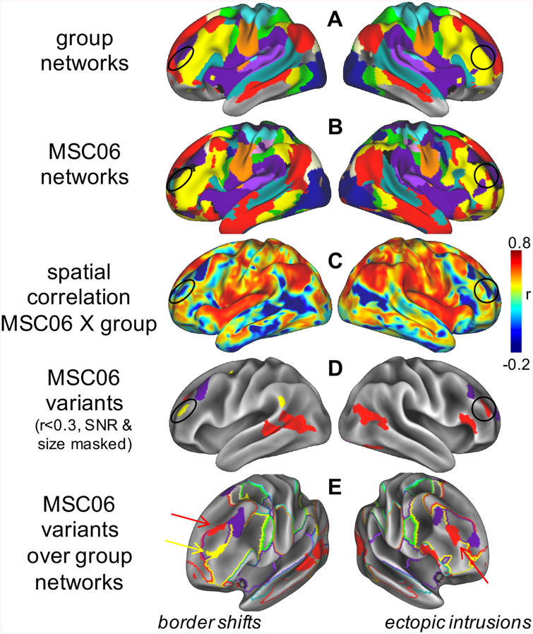
Figure 3: Network variants.
Comparison of (A) group networks and (B) an individual from the MSC (MSC06, colors match Fig. 1). (C) Some locations exhibit low similarity to the group (a few examples are circled), which can be identified through vertex-wise spatial correlation; (D) we call these low-similarity locations network variants. (E) Variants may represent shifts of network borders (left) or isolated ectopic intrusions (right). We hypothesize that network variants are caused by stable factors that reprioritize the neural functions of cortical areas, causing shifts in the boundaries of cortical networks and ectopic intrusions. These altered prioritizations lead to changes in the dominant systems-level relationships of a region (e.g., increasing FC to relevant regions in alternate networks), causing these regions to appear as network variants. Thus, network variants may be related altered brain function during tasks and behavioral responses across individuals. See (91).

