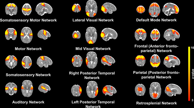
Fig. 1. Resting State Networks in the Neonatal Brain.
Coronal, axial and sagittal examples of 12 independent components extracted with probabilistic ICA are overlaid on a neonatal term-equivalent template. The spatial representation of the RSN was threshold to a z-statistic between 3 and 10. The 12 ICA networks depicted correspond to somatosensory/motor (paracentral), motor, somatosensory, auditory, lateral visual, mid visual, left and right posterior temporal (temporo-parietal), default mode network (DMN), Frontal (anterior fronto-parietal), Parietal (posterior fronto-parietal) and retrosplenial RSNs.

