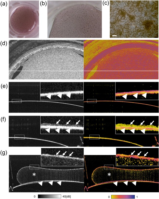
Figure 1.
In-vitro observation of human foetal RPEs and HEK293T cells devoid of melanin by PS-OCT. Digital photograph (a), stereomicroscopic image (b), and microscopic image (c) of a foetal human retinal pigment epithelial (hRPE) sheet in a transwell presenting a cobblestone-like appearance with pigmented cells. The sheet was used after 71 days of seeding in a transwell. En face map of polarization-sensitive optical coherence tomography (PS-OCT) intensity imaging (left) and entropy imaging(right) of the hRPE sheet (d). PS-OCT intensity (left) and entropy (right) of cross-sectional B-scan images of the transwell membrane only (e), human embryonic kidney (HEK) 293T cells on the membrane as a negative control (no melanin containing cells) (f), and the hRPE sheet on collagen gel on the membrane (g). In (d–f), arrowheads indicate a transwell membrane with high intensity and high entropy; arrows indicate presence of cells; asterisks indicate collagen gel. Scale bar, 50 µm.

