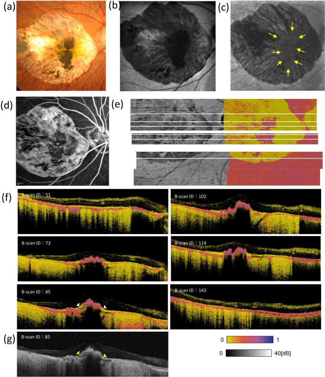
Figure 5.
Ophthalmologic and PS-OCT images of the patient that received hiPSC-RPE sheet transplantation. Colour fundus photograph (a), fundus autofluorescence (AF) images (b,c), and fluorescein angiography image (d) of an eye with transplanted autologous human induced pluripotent stem cell-derived retinal pigment epithelial (hiPSC-PRE) cell sheet. AF images were taken at 532 (b) and 787 (c) nm. Polarization-sensitive optical coherence tomography intensity (right) and en face entropy (left) maps of the eye with transplanted hiPSC-PRE (e); six lines of cross-sectional B-scan entropy images (f); one line of cross-sectional intensity B-scan image (g).

