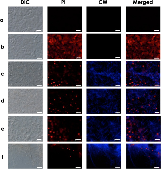
Fig. 5.
Microscopic analyses showing the mycelial trap covering the monolayer formed by lung epithelial cells. A549 cells were cultured in the absence (a) or in the presence of conidia of S. apiospermum (b), S. minutisporum (c), S. aurantiacum (d), and L. prolificans (e) after 24 h. Subsequently, the interaction systems were incubated with Calcofluor white (CW), to localize the mycelial cells, and propidium iodide (PI), to evidence the A549 plasma membrane injury. Note that after 24 h of co-culturing of fungal conidia and A549 cells, only mycelia were observed using phase-contrast (DIC) and fluorescence microscopy. Bars, 50 μm

