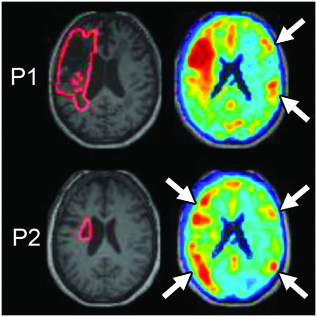
Figure 4.

Functional anomaly maps demonstrate abnormal function distant from lesions, consistent with diaschisis (adapted from DeMarco & Turkeltaub, 2018a). Machine learning analysis of resting-state functional magnetic resonance imaging data is used to grade the degree to which spontaneous brain activity differs in individual stroke survivors from a group of controls. Maps are shown for two individuals (P1 and P2). P1 has a large cortical lesion, and dysfunction is identified at the site of the anatomical lesion and also in regions opposite the stroke in the right hemisphere (white arrows). P2 has a subcortical lesion with widespread cortical dysfunction in the lesioned hemisphere and in locations of the right hemisphere opposite areas of prominent left hemisphere dysfunction (white arrows).
