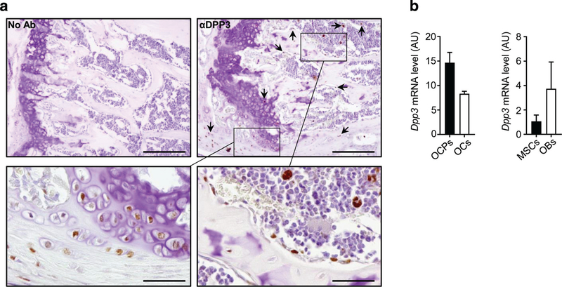
Fig. 1.
DPP3 is expressed in the bone tissue. (A) Representative images of immunohistochemical analysis of DPP3 expression in the femur of WT C57BL/6J mouse. Upper left, negative control; upper right and lower left and right, staining with mouse antibody αDPP3 at different magnification. Scale bars = upper panels 200 μm; lower panels 50 μm. Arrows indicate representative positive cells. (B) qPCR for murine Dpp3 expression in in vitro osteoclastogenesis (n = 3) and osteoblastogenesis (n = 2). In the latter, bars indicate the range. For each evaluation, n ≥ 2. DPP3 = dipeptidyl peptidase 3; OCP = osteoclast precursor; OC = osteoclast; MSC = mesenchymal stromal cell; OB = osteoblast.

