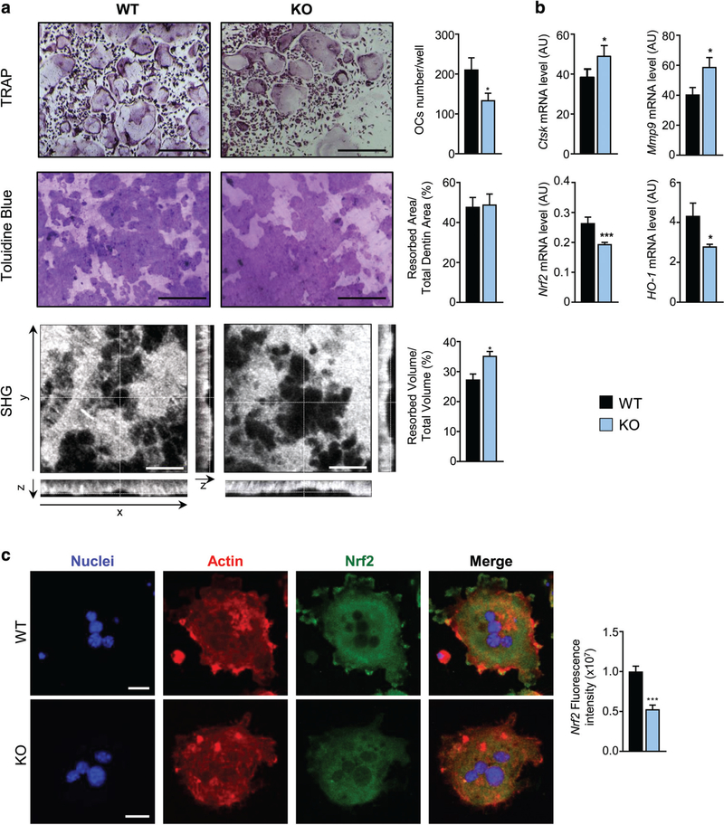
Fig. 6.
Loss of DPP3 increases in vitro osteoclast resorption activity and impairs Nrf2 signaling. (A) Upper panels: representative images of TRAP-stained in vitro differentiated osteoclasts from WT and Dpp3 KO osteoclast precursors. Middle panels: Toluidine blue–stained resorption pits on dentin. Scale bar = 400 μm. Lower panels: SHG of dentin collagen in the different projections. Scale bar = 40 μm. Graphs on the right represent quantification analysis for each evaluation. (B) qPCR analysis of osteoclast functional genes and Nrf2 pathway in in vitro–differentiated WT and Dpp3 KO osteoclasts. For each evaluation, n ≥ 6 per genotype. (C) Representative images of immunofluorescence analysis of in vitro–differentiated WT and Dpp3 KO osteoclasts stained as indicated, and Nrf2 fluorescence intensity. *p < .05, ***p < .001. SHG = second harmonic generation.

