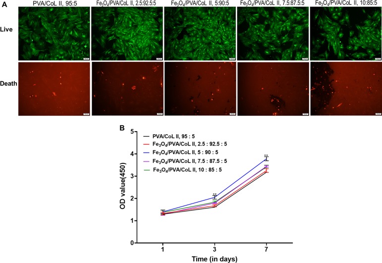
Figure 7.
(A) Staining for living and dead cells on magnetic nanocomposite hydrogels with different Fe3O4 contents after 7 days of cell culture. Cells were inoculated in samples from each group; after inoculation, all cells proliferated well. The cell proliferation of the Fe3O4/PVA/COLII,5:90:5 group was better than that of the other groups. (B) CCK-8 cellular proliferation results from magnetic nanocomposite hydrogels with different Fe3O4 contents. Cells were inoculated on each group, survived, and proliferated. On the third day after inoculation, the cell proliferation of Fe3O4/PVA/COLII,5:90:5 was notably better than the other tested groups. This difference was significant until the 7th day after inoculation (*P < 0.05, **P < 0.01).

