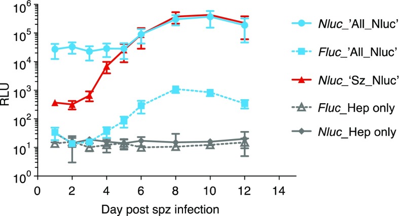
Figure 2.
Total Firefly luciferase (Fluc) and Nanoluc (Nluc) bioluminescence signals measured in the P. cynomolgi dual reporter (‘All_Nluc’) reporter line (blue) and the single reporter (‘Sz_Nluc’) control line (red) at different time points post sporozoite infection of primary rhesus hepatocytes. Results are expressed as average Relative Light Units (RLU) ± s.d. of three independent infections in 96-well plates (triplicate wells each). For reference, background Fluc and Nluc counts are depicted of uninfected hepatocytes from one experiment (gray lines, triplicate wells ± s.d.).

