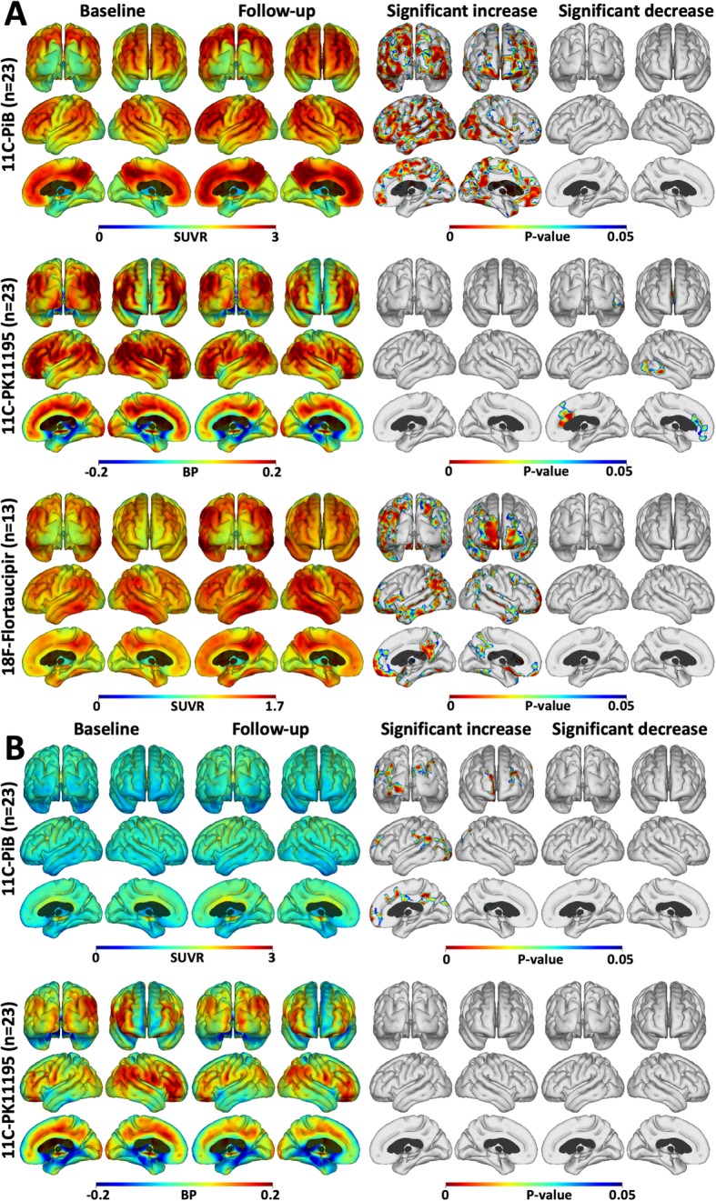
Fig. 1.

Cortical surface maps of mean tracer uptake and changes. a Cortical surface maps of mean PiB (upper), PK (middle) and flortaucipir (lower row) uptake for the high PiB MCI subjects at baseline and after 2 years follow-up. On the right, results of a paired t test between baseline and follow-up show increased amyloid in all cortical association areas, increased tau in frontal and occipital cortical areas, and reduced inflammation in fronto-temporal cortical regions after 2 years. b Mean PiB (upper) and PK (lower) uptake for the low PiB subjects at baseline and after 2 years follow-up. The results of a paired t test show small areas of increased amyloid, but no changes in inflammation levels over 2 years (P < 0.05; cluster FWE rate, P < 0.05)
