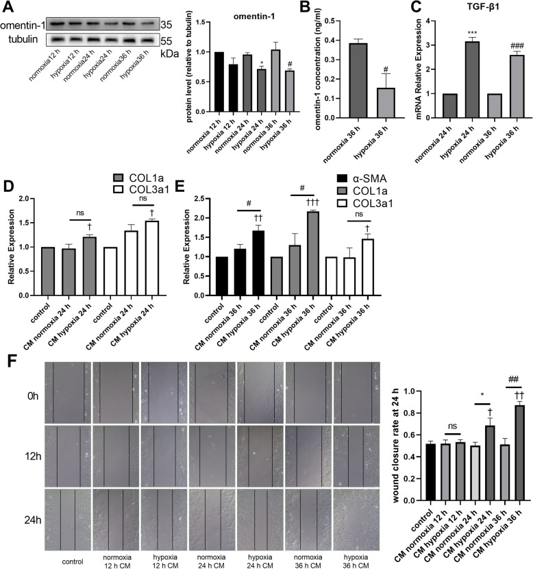
Fig. 4.
Hypoxia resulted in a change in the secretion phenotype of adipocytes. Omentin-1 protein levels in adipocytes was detected via western blotting (a). The concentrations of omentin-1 in the CM of adipocyte treated with normoxia or hypoxia were determined by ELISA (b). The mRNA levels of TGF-β1 in adipocytes (24 h normoxia, 24 h hypoxia, 36 h normoxia, 36 h hypoxia) was detected via RT-qPCR. Expression of mRNA was normalized to GAPDH. (c) The mRNA levels of α-SMA, COL1a, and COL3a1 in non-CM-treated or CM-treated CFs were detected via RT-qPCR. The mRNA level was normalized to GAPDH. (d, e) (f) Representative images of CFs scratch assay (100× magnification). The values were mean ± SEM of three independent experiments. *P < 0.05 vs normoxia 24 h group, ***P < 0.001 vs normoxia 24 h group, #P < 0.05 vs normoxia 36 h group, ###P < 0.001 vs normoxia 36 h group, †P < 0.05 vs control group, ††P < 0.01 vs control group, †††P < 0.001 vs control group

