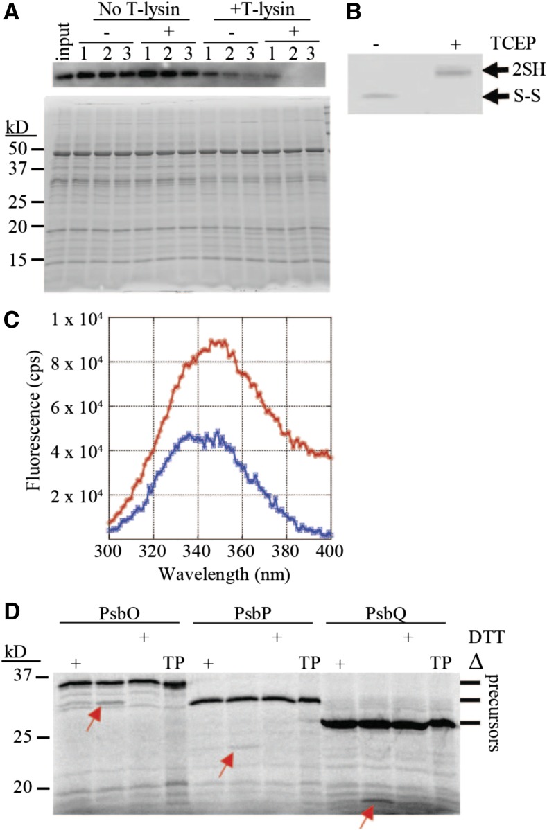
Figure 1.
Disulfide Bond Reduction Alters the Structure of Plsp1.
(A) Proteins extracted from pea thylakoids with 0.25% v/v Triton X-100 were pretreated with (+) or without (-) 10 mM TCEP followed by incubation with or without thermolysin. Aliquots taken at 10 (1), 20 (2), and 30 (3) min were analyzed by SDS-PAGE and immunoblotting with an antibody against Pea Plsp1 (top panel). A second gel was loaded with half of the amount of each sample and stained with Coomassie Brilliant Blue after electrophoresis (bottom panel). Input = untreated thylakoid extracts.
(B) An aliquot of the +/− TCEP pretreated thylakoid extracts was analyzed by nonreducing SDS-PAGE and immunoblotting with the antibody against Pea Plsp1. 2SH = reduced Plsp1; S-S = oxidized Plsp1.
(C) Emission spectrum of purified T7-Plsp171-291 (∼0.1 μM) in 1% w/v octyl glucoside after pretreatment with (Blue) or without (Red) 50 mM DTT plotted as fluorescence in counts per second (cps) versus wavelength. Shown are the spectra after subtracting those of buffer blanks. The excitation wavelength was 285 nm.
(D) Processing activity of purified T7-Plsp1 against prPsbO, prPsbP, or prPsbQ. T7-Plsp1 used in (C) was mixed with 35S-Met-labeled substrates after being boiled for 10 min (Δ) or pretreated with 50 mM DTT on ice for 20 min. Reaction mixtures were incubated at ∼25°C for 30 min and analyzed by SDS-PAGE and autoradiography. Arrows denote the processed forms of each substrate tested. TP = translation products.

