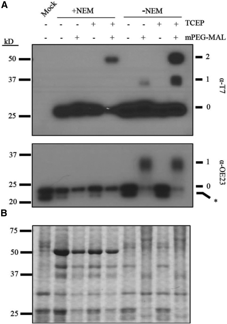
Figure 2.
Plsp1 Cys Form a Disulfide Bond in Chloroplasts.
(A) PEG-MAL labeling of Cys in isolated chloroplast membranes. Thiol labeling was performed as described in Methods. Samples were analyzed by SDS-PAGE and immunoblotting using the α-T7 antibody for detection of T7-Plsp1 (top panel) or the α-OE23 antibody as a control (bottom panel). Unlabeled protein (0). 1 or 2 Cys residues labeled with mPEG-MAL, (1) and (2), respectively. Asterisk indicates unknown immunoreactive protein in N. benthamiana chloroplast membranes.
(B) Coomassie-stained gel of the same samples analyzed in (A).

