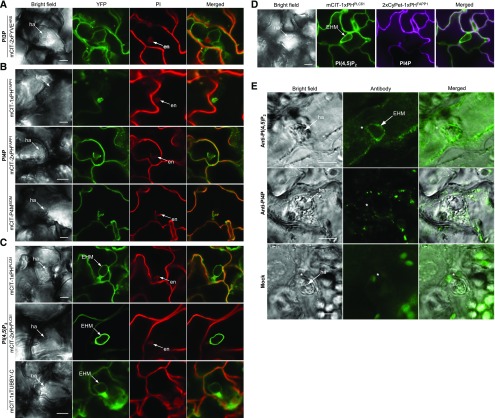
Figure 1.
Differential Targeting of Phosphoinositides to the Haustorial Periphery of the Powdery Mildew Ec.
(A) to (C) Leaves of Arabidopsis plants expressing biosensors were inoculated with Ec and viewed with a confocal microscope at 2 DAI. Fungal structures and plant cell walls were stained with propidium iodide (PI). en, encasement; ha, haustorium. Bars = 10 µm.
(A) Representative images of PI3P biosensor mCIT-2xFYVEHRS.
(B) Representative images of PI4P biosensors mCIT-1xPHFAPP1, mCIT-2xPHFAPP1, and mCIT-P4MSiDM.
(C) Representative images of PI(4,5)P2 biosensors mCIT-1xPHPLCδ1, mCIT-2xPHPLCδ1, and mCIT-1xTUBBY-C.
(D) Simultaneous labeling of PI(4,5)P2 (mCIT-1xPHPLCδ1) and PI4P (2xCyPet-1xPHFAPP1) during haustorium formation at 2 DAI. Bar = 10 µm.
(E) Immunofluorescence of Ec-infected leaf epidermal cells with the antibodies to PI(4,5)P2 [anti-PI(4,5)P2] and PI4P (anti-PI4P). Distribution of PI(4,5)P2 and PI4P in Ec-infected cells was revealed by whole-mount immunolocalization of leaf epidermal tissues of Arabidopsis plants at 2 DAI. Images of mock were taken in the absence of primary antibody. Asterisks indicate Ec penetration sites in epidermal cells. Bars = 10 µm.

