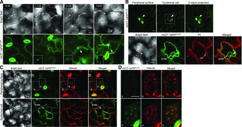
Figure 4.
Induced PI(4,5)P2 Dynamics in Host Cells in Response to Powdery Mildew Infection.
(A) Time-course responses of PI(4,5)P2 dynamics revealed by the mCIT-1xPHPLCδ1 probe in Ec-infected epidermal cells at 9 to 14 hpi. Notably, signals of mCIT-1xPHPLCδ1 were focally accumulated underneath the penetration site initially at ∼11 hpi and then targeted the EHM during haustorial development. Asterisks indicate the penetration sites that are enlarged in insets for close views; arrowheads indicate the EHM.
(B) Enhanced production of PI(4,5)P2 specifically in Ec-colonized cells. The bottom row shows enlarged views of an Ec-colonized cell at 24 hpi, showing enhanced PI(4,5)P2 signals at the EHM as well as along the PM of the infected cell. Fungal structures and plant cell walls were stained with PI. Induced accumulation was observed in 47 of 79 Ec-colonized cells.
(C) and (D) Association of induced PI(4,5)P2 production with PM and endocytic processes in Ec-colonized cells. Ec-inoculated leaves at 24 hpi were incubated in FM4-64 for 15 min.
(C) An Ec-infected cell (a) and a neighboring noninfected cell (b) are highlighted in dash-lined boxes. The same inoculation sites were viewed on the peripheral surface (top) or inside the cell (bottom) of leaf epidermis.
(D) Enlarged views of an Ec-infected cell (a) and a noninfected cell (b). Note that PI(4,5)P2 signal revealed by mCIT-1xPHPLCδ1 was induced only in the Ec-colonized cell and colocalized with FM4-64-labeled endocytic PM compartments on the peripheral surface of the infected cell.
app, appressorium; c, conidium; en, encasement; ha, haustorium. Bars = 10 µm.

