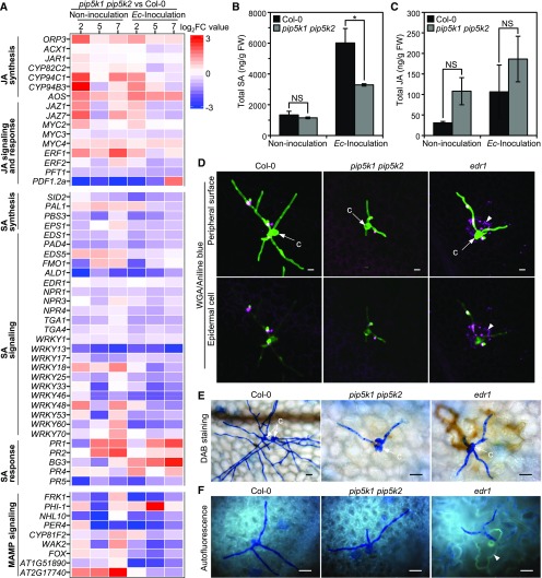
Figure 6.
Defense Responses in pip5k1 pip5k2 Mutants against Powdery Mildew Infection.
(A) Transcriptomic profiling of differentially expressed genes in SA and JA biosynthesis, signaling and response pathways, and MAMP signaling between pip5k1 pip5k2 mutant and Col-0 plants without or with Ec inoculation at 2, 5, and 7 DAI. Heat maps display log2 fold change (log2FC) values for pairwise comparison between the pip5k1 pip5k2 mutant and Col-0 at each time point.
(B) and (C) Levels of SA and JA in Col-0 and the pip5k1 pip5k2 mutant. Total amounts of SA (B) and JA (C) were measured in leaf tissues without or with Ec inoculation at 5 DAI. Data are means ± sd (n = 3 biological replicates). *, P < 0.05; NS, no significant difference, Student’s t test. FW, fresh weight.
(D) to (F) Detection of callose deposition, H2O2 accumulation, and autofluorescence material production in Ec-infected Col-0, pip5k1 pip5k2, and edr1 plants at 48 hpi. Arrowheads indicate cell death in the edr1 mutant accompanied by callose deposition, H2O2 accumulation, and autofluorescence. c, conidia. Bars = 20 μm.
(D) Callose deposition. Ec-inoculated leaves were fixed and stained by both aniline blue and Alexa Fluor 488-conjugated wheat germ agglutinin (WGA). The images were obtained by merging the confocal optical sections (Z-stacks).
(E) H2O2 production. Ec-inoculated fresh leaves were stained by 3,3'-diaminobenzidine, fixed, and viewed by compound microscopy. H2O2 accumulation is indicated by brownish color.
(F) Accumulation of autofluorescence materials. Ec-inoculated leaves were fixed, and the autofluorescence was directly viewed by fluorescence microscopy.

