Figure 8.
Regulation of PI(4,5)P2 Controls Disease Development in Plants and the Lifestyle of the Hemibiotrophic Fungal Pathogen Ch.
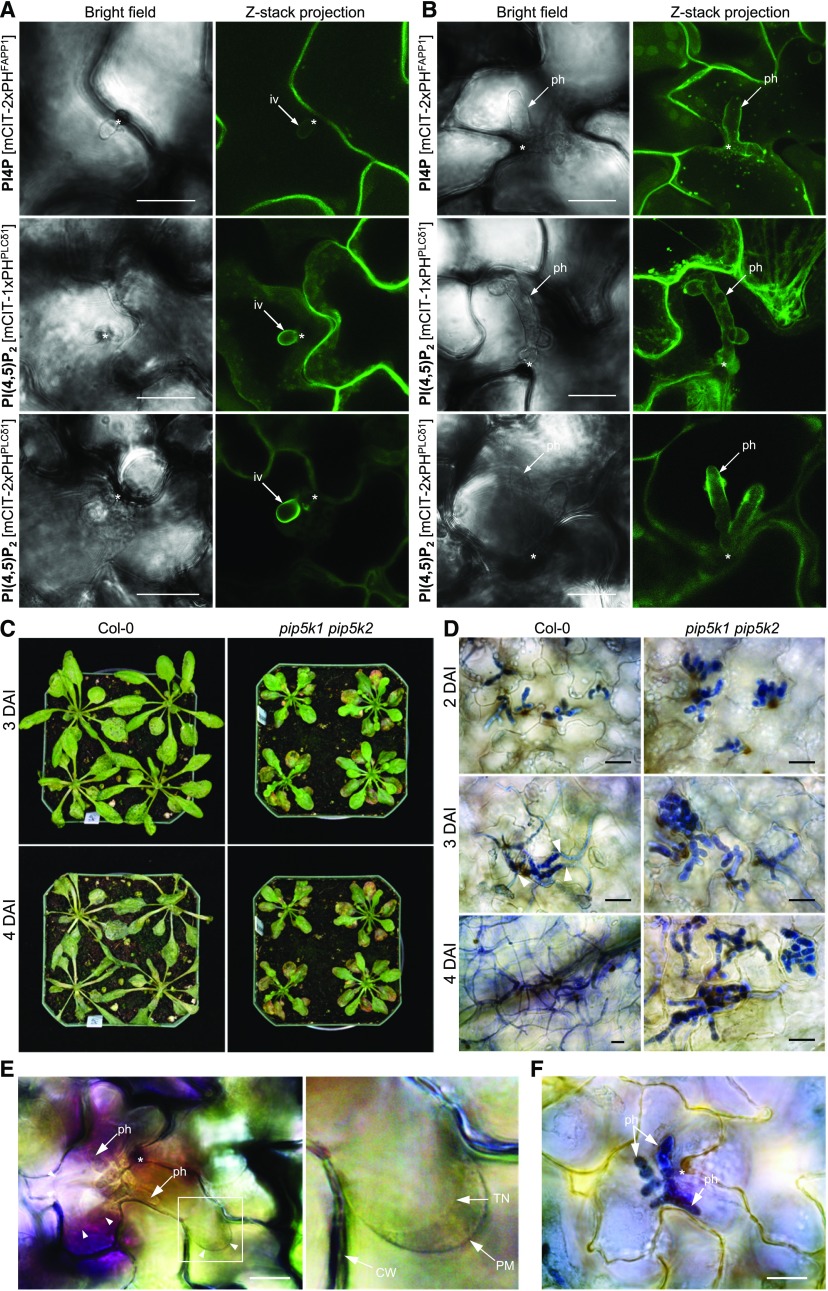
(A) and (B) Association of the PI4P biosensor mCIT-2xPHFAPP1 and the PI(4,5)P2 biosensors mCIT-1xPHPLCδ1 and mCIT-2xPHPLCδ1 with the biotrophic stages of the Ch life cycle. Both PI4P and PI(4,5)P2 signals targeted the surface of infection vesicles (iv; [A]) and primary hyphae (ph; [B]). Asterisks indicate the penetration sites.
(C) Disease development on Col-0 and pip5k1 pip5k2 plants. Ch-inoculated plants were photographed at 3 and 4 DAI.
(D) Microscopic images of Ch-infected leaf tissues. In pip5k1 pip5k2 leaves, extensive bulbous primary hyphae were restricted within the first infected epidermal cells during the infection time course 2 to 4 DAI, whereas in Col-0 plants, thin necrotrophic hyphae developed at 3 DAI and rapidly spread into neighboring cells. Infected leaf tissues were stained with trypan blue.
(E) and (F) Extended biotrophic stages of Ch infection in the pip5k1 pip5k2 mutant.
(E) Viability of Ch-infected cells at 4 DAI is shown by the host protoplasm contracting from the cell wall (CW) after plasmolysis. The right panel shows an enlarged view of the boxed area in which tonoplast (TN) is clearly distinguishable from the PM.
(F) Leaf sample showing that the same Ec-infected site in (E) was fixed and stained for fungal hyphae with trypan blue.
Bars = 20 μm.

