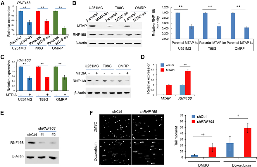
Figure 3. RNF168 Expression Is Attenuated in MTAP-Deficient GBM Cells.
(A and B) The expression of RNF168 was determined in the parental U251MG cell line and its derivative MTAP-ko cell line by (A) qRT-PCR and (B) immunoblotting (the quantification of the relative abundance of RNF168 in each lane is shown in the right panel).
(C) GBM cell lines were treated with DMSO or MTDIA (3 μM) for 24 h, and the expression of RNF168 was determined by qRT-PCR (left panel) and by immunoblotting (right panel).
(D) The RNF168 transcript level in a patient-derived, naturally MTAP null GBM cell line (GBM #13–0302), without or with exogenous MTAP restored (via retroviral delivery, denoted MTAP+), was determined by qRT-PCR (note that no MTAP transcript was detected in the “vector” line).
(E) Immunoblot detection of RNF168 in U251MG cells without (shCtrl) or with knockdown of RNF168 (shRNF168).
(F) U251MG cell lines, without or with RNF168 knockdown, were treated with vehicle control (DMSO) or with doxorubicin (0.1 μM, 24 h), and an alkaline comet assay was performed. Quantification of the tail moment is shown in the right panel.
Student’s t-test was used for statistical analysis, except for tail-moment quantification, where Mann-Whitney U test was used. *p < 0.05, **p < 0.01. Error bars represent mean ± SD.

