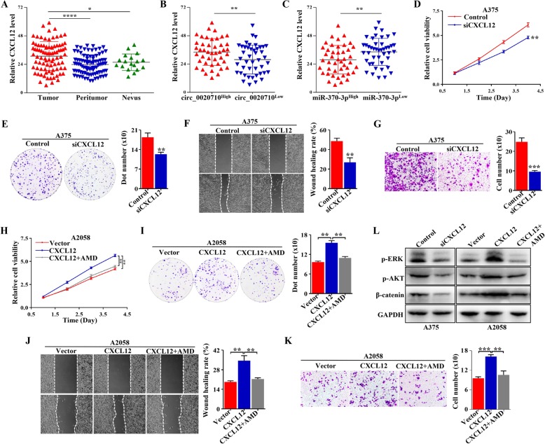
Fig. 5.
Elevated CXCL12 promotes melanoma progression. a IHC assay was used to detect the expression of CXLC12 in 88 paired melanoma and normal tissues and 18 benign nevi tissues. b Relative CXCL12 expression in different melanoma samples according to the expression of circ_0020710. c Relative CXCL12 expression in different melanoma samples according to the expression of miR-370-3p. d-g Colony formation, wound healing, transwell invasion, and CCK-8 assays performed in A375-Control and A375-siCXCL12 cells. h-k CCK-8, Colony formation, transwell invasion and wound healing assays performed in A2058 cells treated with Vector, CXCL12, and CXCL12 + AMD3100. l Western blot assay was used to detect the p-ERK, p-AKT, and β-catenin levels in melanoma cells with different treatment, GAPDH was used as a negative control. Paired student’s-t test, unpaired student’s t-test, Mann-Whitney U test, one-way ANOVA test and Kruskal-Wallis test were used for the statistical analyses. *p < 0.05; **p < 0.01; ***p < 0.001; ****p < 0.0001; ns, no significant

