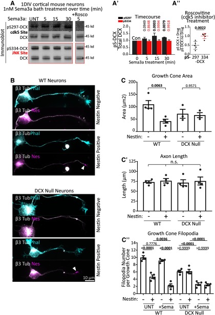
Figure 10.
DCX is a cdk5 substrate downstream of Sema3a, and Dcx-null neurons respond in a nestin-independent manner. A, Sema3a treatment of 1 DIV cultured mouse cortical E16 neurons significantly increases DCX phosphorylation at S297, but not at S334. Rightmost lane, Neurons were treated with 1 nm Sema3a for 5 min with preincubation of the cells with 10 μm roscovitine for 30 min. The inhibitor lane was from the same blot as the other lanes, but separated by intervening lanes that were removed. A′, Quantification of Sema3a time course. A, Blots were subjected to densitometry analysis, and the ratio of phospho-DCX signal relative to the total DCX is plotted. DCX phosphorylation at pS297, but not at pS334, begins quickly after Sema3a stimulation and becomes significant at 15 and 30 min. Error bars indicate SD. Statistical analysis was performed at each time point compared with untreated (UNT) with an ordinary one-way ANOVA with Dunnett's correction. n = 5 independent experiments. p values of <0.05 are indicated in bold. A′′, Quantification of roscovitine sensitivity (A, rightmost lane). Each value was obtained from densitometry analysis of Western blots, where the relative level of p-DCX was compared with total DCX. The values in the plot are a ratio of the relative phosphorylation after inhibitor (drug) treatment compared with no inhibitor treatment. Error bars indicate SD. Statistical analysis was performed with a two-tailed unpaired t test. N = 5 independent experiments. B, Representative images of nestin-positive or -negative individual neurons from WT or Dcx-null primary E16 cortical neuron cultures 1 DIV (Stage 3) quantified in C and C′. Top image for each condition represents phalloidin staining. Bottom image for each condition represents nestin staining of the same neuron. Arrowheads indicate Nestin-positive GCs. Top, bottom, β3 tubulin shown to both visualize the cells overall morphology and to confirm neuronal identity. C, C′, C′′, Quantification of morphologic characteristics of nestin-negative and -positive neurons derived from WT or Dcx-null E16 cortex at 1 DIV (Stage 3). WT cells show nestin-dependent decrease in GC size, whereas Dcx-null neurons do not. Axon length was not significantly different in any condition. When treated with 1 nm Sema3a for 5 min, filopodial retraction in WT neurons was dependent on nestin expression. In Dcx-null neurons, there was a filopodia decrease after Sema3a treatment in nestin-positive and -negative neurons (independent of nestin). C, Statistical analysis was performed with one-way ANOVA with Sidak's multiple comparison correction (with pairs tested shown). C′, Statistical analysis was performed with one-way ANOVA with Tukey's multiple comparison correction. C′′, Statistical analysis was performed with one-way ANOVA with Sidak's multiple comparison correction (with pairs tested shown). C, C′, C′′, n = 4 or 5 independent experiments with 20-30 cells counted in each set. p values of <0.05 are indicated in bold. Error bars indicate SEM.

