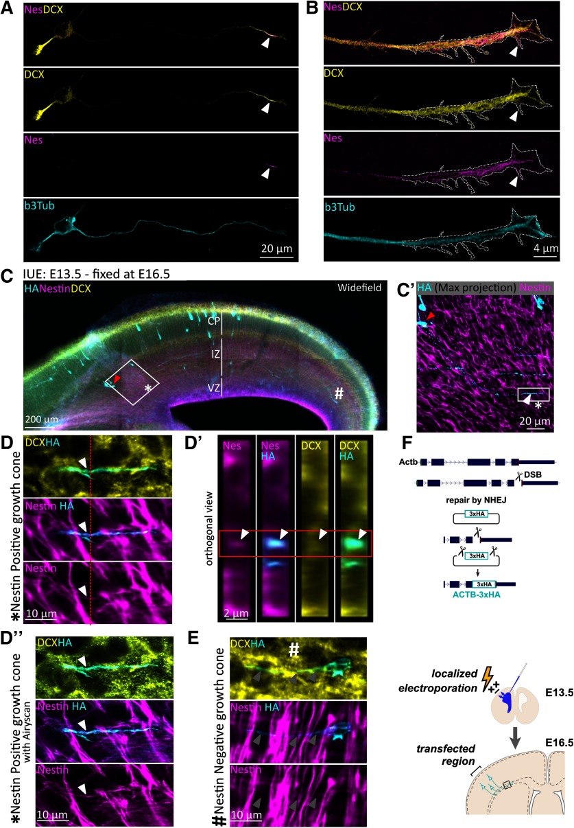
Figure 5.
Nestin colocalizes with DCX in axons of newly born neurons in vitro and in vivo. A, Confocal images of an E16 cortical mouse Stage 3 neuron after 1 DIV culture, counterstained for endogenous DCX, nestin, and β3 tubulin. DCX is enriched at the distal region of all neurites, whereas nestin immunostaining labels only the distal region of the growing axon (arrowhead), but not of the minor neurites. B, Enhanced resolution confocal imaging of nestin in the distal region of an axon together with DCX. Distally, some nestin filaments appear to extend into the central region of the GC and into some proximal filopodia (arrowhead). C, Wide-field immunofluorescence image of a coronal section of E16.5 mouse cortex to illustrate the sparse labeling of individual neurons achieved with knockin of HA-tag using CRISPR and IUE. In all panels, red arrowhead indicates the cell body that projects the HA/Nes/DCX-positive GC. *Nestin-positive GC. #Nestin-negative neuron (shown in D′′, E). The major cortical layers are labeled: VZ, Ventricular zone. Antibody labeling is indicated in each panel. C′, Inset of square in C demonstrating HA staining of a single Stage 3 neuron with the entirety of its axon imaged in a confocal stack (maximum projection). White arrowhead indicates the distal axon/GC. Red arrowhead indicates the cell body. D, D′, Confocal images. D′′, Airyscan-enhanced resolution image. D, High-magnification zoom of the inset square of C′ of the GC marked with * in C and C′. A single z plane is shown. D′, Orthogonal projection of the region indicated in the red dashed line of D. Red box outlines the relevant HA-labeled cell, which is positive for nestin and DCX. D′′, High-magnification Zeiss Airyscan confocal image equivalent of D. E, High-magnification zoom of the GC marked with # in C. Arrowheads point along the length of the axon; nestin is not detected in this GC. F, Diagram of the experimental strategy to detect nestin and DCX in single GCs in mouse cortex. E13.5 mouse cortex was electroporated in utero to sparsely introduce three HA tags into β-actin (Actb-3xHA) by CRISPR. Brains were fixed at E16.5. The CRISPR strategy is depicted diagrammatically.

