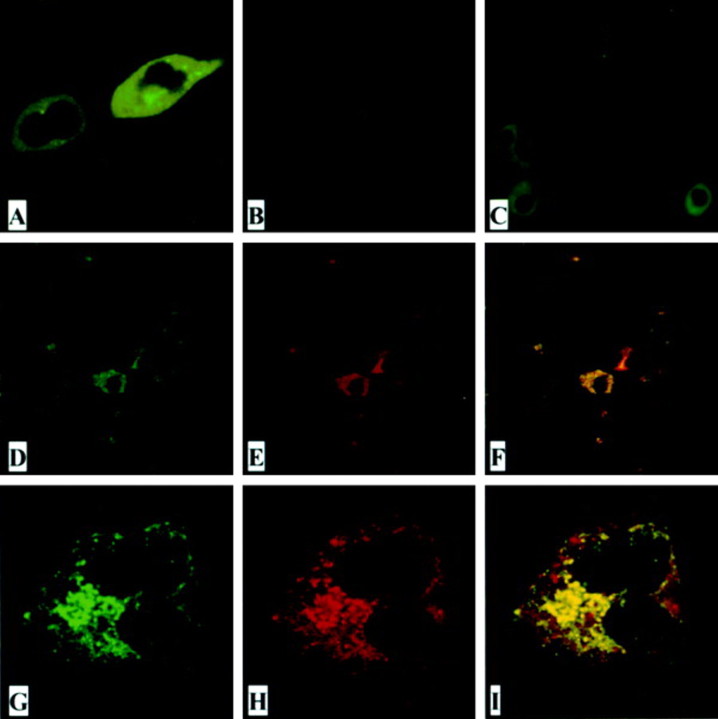
Figure 4.
The subcellular localization of open reading frame (ORF)-8a. A, HEK 293T cells transfected with plasmid pORF8a-EGFP. B, Severe acute respiratory syndrome coronavirus (SARS-CoV)-infected VeroE6 cells immunostained with preimmunized rabbit serum. C, SARS-CoV-infected VeroE6 cells immunostained with rabbit anti-ORF8a antiserum R26 (1:100 dilution). D-I, VeroE6 cells cotransfected with a mitochondrial expression plasmid (pEF-CKMt2-green fluorescent protein [GFP]) and pHA-ORF8a and immunostained with rabbit anti-ORF8a antiserum R26 (1:500 dilution). Rhodamine-conjugated goat anti-rabbit antibodies were used as a secondary antibody in the immunofluorescent antibody assay test. Green represents GFP (D and G); red represents ORF8a protein (E and H). Yellowish areas in merged panels (F and I) indicate ORF8a localized in mitochondria. D-F, Low magnification; G-I, high magnification.

