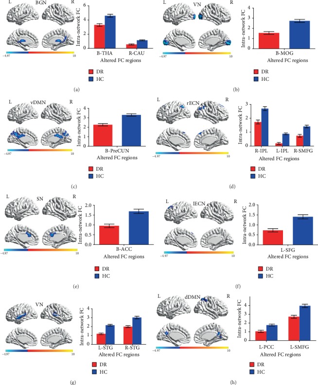
Figure 2.

Brain regions with significant differences for eight RSNs in the DR group vs. the HC group (two-tailed, voxel-level P < 0.01; GRF correction, cluster-level P < 0.05). Cool colors indicated the decreased intranetwork functional connectivity FC in the DR group compared with the HC group, as shown by BrainNet Viewer. (a–h) correspond to different resting-state networks. BGN, VN, vDMN, rECN, SN, lECN, AN, and dDMN. Abbreviations: DR: diabetic retinopathy; HC: healthy control; BGN: basal ganglia network; VN: visual network; vDMN: ventral default mode network; rECN: right executive control network; SN: salience network; lECN: left executive control network; AN: auditory network; dDMN: dorsal default mode network; RSN: resting-state networks; THA: thalamus; CAU: caudate; MOG: middle occipital gyrus; PreCUN: precuneus; IPL: inferior parietal lobule; SMFG: superior medial frontal gyrus; ACC: anterior cingulate gyrus; SFG: superior frontal gyrus; STG: superior temporal gyrus; PCC: posterior cingulate gyrus; GRF: Gaussian random field; L: left; R: right; B: bilateral.
