
Figure 3.
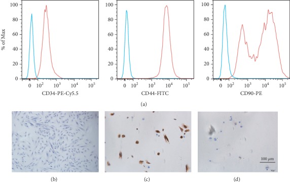
Typical characteristics of hEndoSCs. (a) Flow cytometry detection of the immunophenotypes of hEndoSCs. The cells were positive for CD34, CD44, and CD90. (b) HE staining showed morphology of hEndoSCs. (c) Immunohistochemical identification of vimentin expression. (d) Immunohistochemical identification of keratin expression.