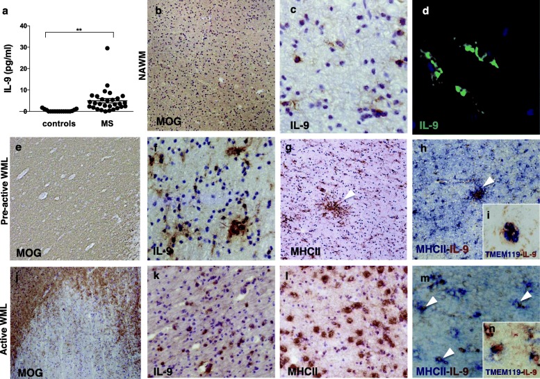
Fig. 1.
IL-9 is expressed in the central nervous system of secondary progressive MS patients. Molecular and neuropathological analysis of IL-9 expression in post-mortem MS white matter. CSF levels of IL-9 in the 29 post-mortem MS cases and 17 age-matched controls (a) (**p < 0.01). In normal appearing white matter (NAWM) identified by myelin oligodendrocyte glycoprotein (MOG) immunostaining (b), rare and scattered IL-9+ cells with microglia morphology (c, d) were detected by immunohistochemistry or immunofluorescence. In pre-active white matter lesions (WML) (e), where clusters of MHC class II+ microglia were present (arrowheads in g), increased expression of IL-9 was detected (f). Double immunohistochemistry demonstrated that most of the IL-9 was expressed by clusters of microglia expressing MHC-II (arrowheads in h) or TMEM-119 (i). In active WML, with on-going demyelination (j) and a high density of microglia/macrophages MHC-II+ (l), elevated IL-9 expression was observed (k), often in combination with MHC-II (arrowheads in m) or TMEM-119 (n). Original magnifications: × 100 (b, e, g, h, l, k), × 200 (c, f, j–m), ×400 (d, i, n)

