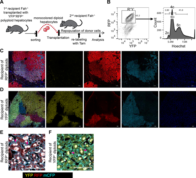
Figure 3. Re-polyploidization of ploidy-reduced cells during liver regeneration.
(A) Experimental scheme for tracing of ploidy-reduced cells in serial recipient mice. (B) Representative FACS plot of a recipient mouse transplanted with ploidy-reduced cells. A mouse serially transplanted with RFP+ monocolored diploid hepatocytes which were originally derived from YFP+RFP+ polyploids is shown. (C, D) Representative microscopic images of the recipient livers repopulated with YFP+ or RFP+ ploidy-reduced 2c cells. Stitched images are shown. (E, F) High-magnification images of (C) and (D). Areas are indicated by dotted line in (C) and (D). Nuclei are shown in white pseudocolor. Scale bars: 100 μm.

