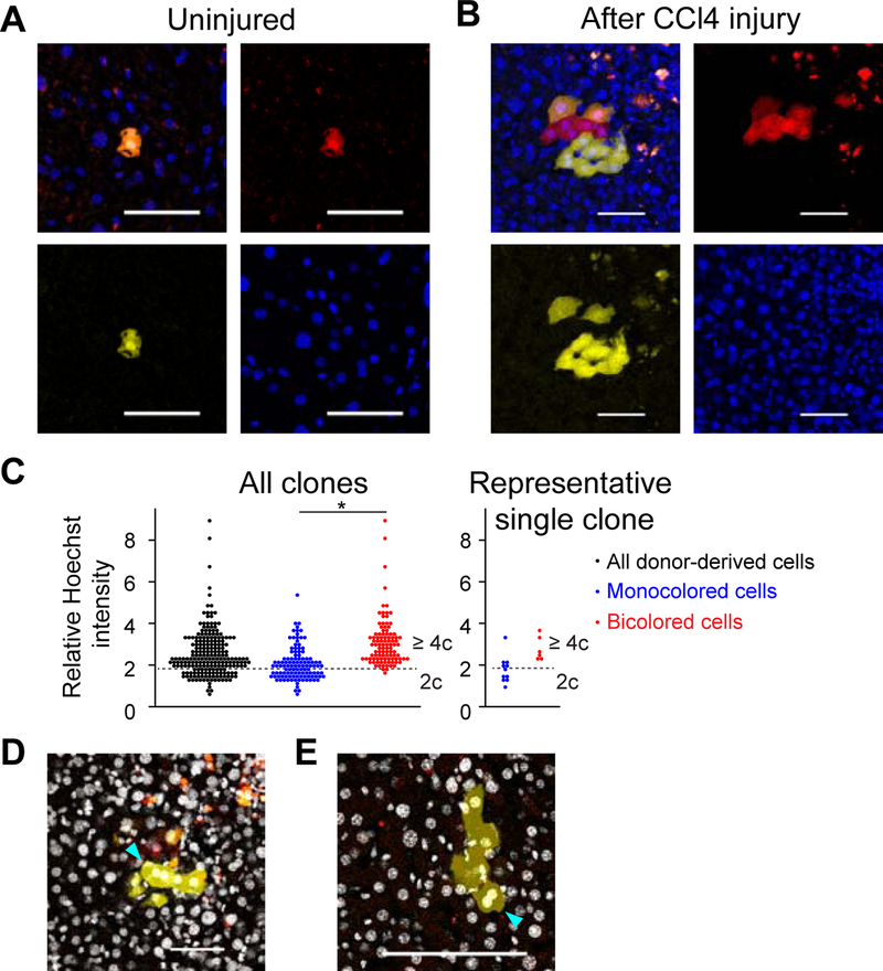
Figure 6. Ploidy reduction and re-polyploidization during CCl4 injury.
(A, B) Representative microscopic images of wild-type mouse livers transplanted with YFP+RFP+ bicolored polyploid hepatocytes. Images of uninjured (A) and CCl4-injured livers (B) are shown. Scale bars: 50 μm. (C) Image cytometry of regenerating clones. Ten clones which contain both bicolored and monocolored cells were analyzed (> 200 cells, 3 mice). Sum of nuclear Hoechst intensity of each donor-derived cell was normalized by the mean Hoechst intensity of the background, and shown as a relative Hoechst intensity. A dotted line that divides polyploid from diploid is set in the same position as in Figure S7A. Result of a representative clone containing ploidy-reduced monocolored cells is shown in the right panel. *p < 0.01 (D, E) Binucleated monocolored cells with ploidy-reduced nuclei. Binucleated cells observed in two clones harboring both bicolored and monocolored cells are indicated by arrowheads. Ploidy of each nucleus in both clones was analyzed in (C). (D) is the same clone as that shown in (B). Nuclei stained with Hoechst are visualized by white pseudocolor. Serial z-stack images are shown in Figures S7B and S7C. The livers were harvested after 6 weeks of CCl4-induced injury. See also Figure S7.

